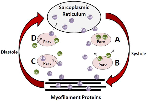
Figure 2.
Working Model for Parv Function in the Heart. A) In the relaxed state, cytosolic Ca2+ is low and Mg2+ is bound to Parv. B) At the beginning of systole, Ca2+ is released from the SR and binds to the myofilament protein cTnC to facilitate contraction. Increased cytosolic Ca2+ concentration causes dissociation of Mg2+ from Parv. C) In diastole, Parv facilitates rapid dissociation of Ca2+ from cTnC by buffering Ca2+ away from the myofilaments, thus facilitating relaxation. D) As Ca2+ is returned to the SR, Ca2+ dissociates from Parv and is replaced by Mg2+.

