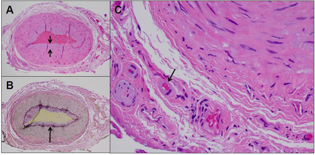
Fig. 2.
Cross-sections of the temporal artery showed mild intimal thickening (short arrows) and an intact internal elastic lamina (long arrow) (A, H&E; B, Van Gieson stain, respectively; magnification 100X). Hyalinazation of vasa vasorum arterioles was noted (C, long arrows), consistent with diabetic vasculopathy. No evidence of inflammation was noted, particularly in the adventitial connective tissue or in association with vasa vasorum or vasa nervosum structures (H&E, magnification 400X. CD45 immunohistochemistry, as previously described [11], confirmed the absence of an inflammatory cell infiltrate (not shown).

