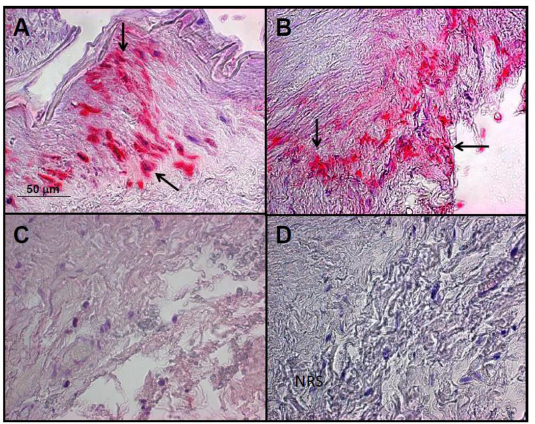
Fig. 3.
The left temporal artery in a patient with multifocal vasculopathy was analyzed for the presence varicella zoster virus (VZV) antigen as previously described [1]. A positive control cadaveric cerebral artery 14 days after VZV infection in vitro (A, pink color, arrows). VZV antigen was seen in the adventitia of the left temporal artery of the subject after staining with anti-VZV antibody (B, pink color, arrows), but not after staining adjacent sections with anti-HSV antibody (C) or normal rabbit serum (D). Magnification 200X.

