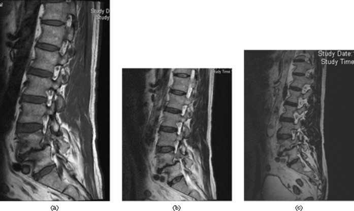
Figure 4.
Pars defect L4. (a) The sagittal T1 weighted and (b) T2 weighted images do not demonstrate the defect well. (c) The defect is best seen on sampling perfection with application-optimised contrasts using different flip angle evolutions (SPACE), sagittal reformat. The SPACE sequence allows for isotropic reconstruction in any plane; the thin slice thickness with still-adequate signal-to-noise ratio allows for better depiction of normal and abnormal anatomy.

