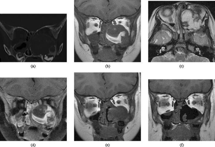
Figure 2.
Case 2. (a) Coronal CT image shows a well-defined, multilobulated soft-tissue mass with diffuse cortical thinning with focal sclerosis. (b) Coronal T1 weighted image shows a mixed-signal lesion with hyperintense signal areas. (c) The lesion has mainly hyperintense signal with hypointense signal rim on the axial T2 weighted image. (d) Coronal contrast-enhanced T1 weighted image with fat saturation shows moderate septa-like contrast enhancement of the lesion. (e) Coronal T1 weighted image shows a recurrent lesion in the primary site. (f) Coronal T1 weighted image shows no evidence of recurrence.

