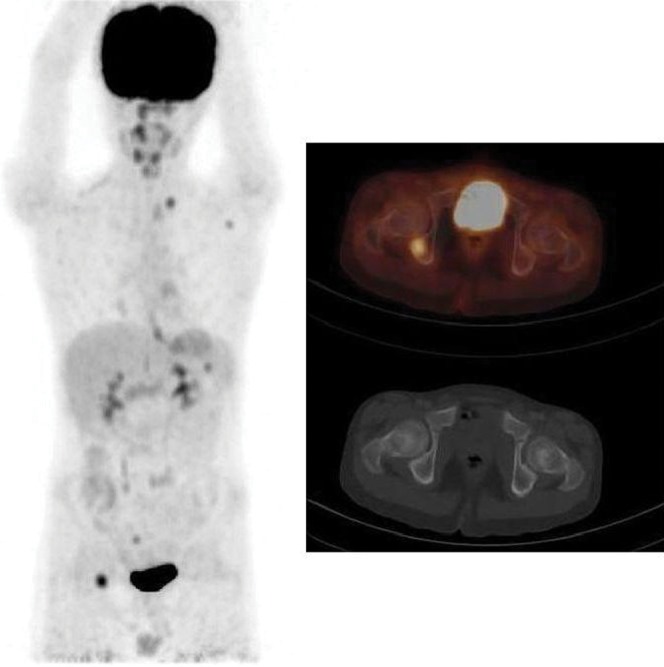
Figure 2.

Fluorine-18-fludeoxyglucose (18F-FDG)-positron emission tomography/CT shows an FDG-avid focus in the right ischium in the maximal intensity projection and transaxial fusion images. The corresponding CT image shows no matching abnormality. FDG-avid disease identified in the left supraclavicular region, the left axilla and the spleen. Bone marrow biopsy was negative in this patient.
