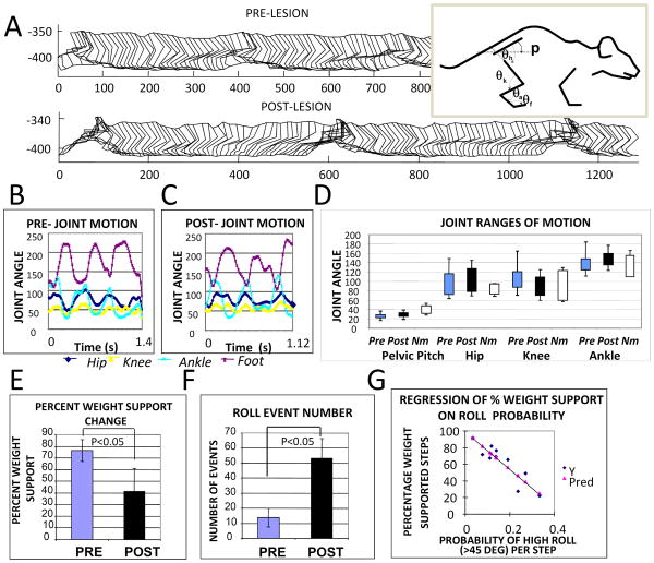
Figure 4.
Kinematics of hindlimbs and pelvis in rats before and after cortical lesions. A. Parasagittal stick figure motion was digitized for multiple step cycles before and after lesion. An example of data from one rat is shown. The measured internal angles are displayed to the right. B Pre-lesion and C Post-lesion joint angles. Pelvic pitch orientation and joint angles of hip, knee, ankle and foot were measured from the captured stick figures pre and post lesion. D. Range of motion for each angles time-series were compared pre-post for 8 WSNST rats (Pre, Post) and for normal rats (Nm). Ranges for data from B/C are shown, together with normal intact rat ranges measured similarly. The maximum and minimum joint angles and their standard deviations in the group of 8 rats tested in detail for pre and post lesion data were compared. In the NST 8 rats tested, statistical comparisons of the kinematic features measured in the parasagittal plane were not significantly different. Neither the ranges of motion, the basic pattern of coordination among joints, nor the period of the hindlimb stepping were significantly altered by the cortical lesions (n=8, p>0.1). E. After lesions, the group percent weight-support decreased significantly (paired t-test, p<0.05). F. After lesion there were increased numbers of pelvic roll events where roll clearly exceeded 45 degrees (paired t-test, p<0.05). The probability of 45 degree roll for each step was calculated from these data and more than doubled post-lesion (paired t-test, p<0.05). The number of non-weight supporting steps in rats was also linearly related to the number of roll events (r2 =0.83, slope coefficient ∼2 and significance p<0.0005). G The percentage of weight-supported steps in rats was negatively correlated to the probability of roll per step (r2=0.81, slope coefficient significance p<0.0001). Thus Hindlimb kinematics were not altered significantly, but pelvic roll was increased and related to quality of weight support. Reworked from Figure 6 in Giszter et al., 2008, J. Neurophysiology, 100(2):839-51, Epub 2008, May 28.31

