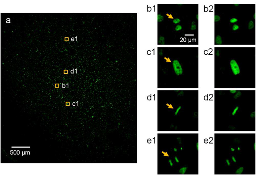
Figure 2.
Wide FOV fluorescence imaging of the GFP cells. (a) The 3.7 mm × 3.5 mm image. (b1, c1, d1, e1) Cropped images of typical cells in (a), including G1 (b1), G2 (c1), metaphase (d1), and anaphase (e1) (arrows). (b2, c2, d2, e2) The same cells as imaged by a conventional microscope with a 20x/0.4 NA objective.

