Fig.6.
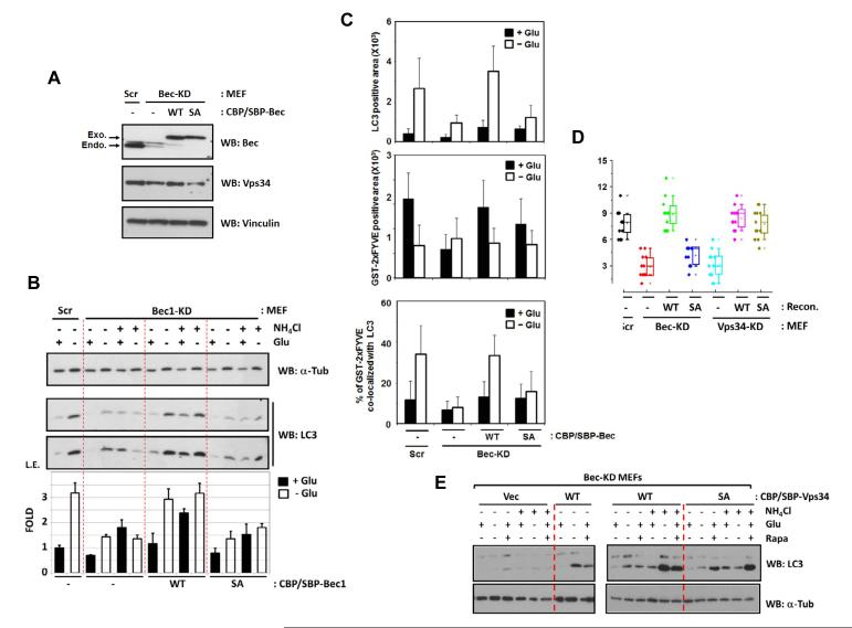
Beclin1 S91/S94 phosphorylation is required for autophagy induction.
(A) Beclin1 expression level in Beclin1 knockdown (KD) and reconstituted MEFs. Scr denotes scramble shRNA control.
(B) Cells expressing the Beclin1 S91/94A mutant are compromised in LC3 lipidation in response to glucose starvation (n=3).
(C) Beclin1 S91/S94A mutant is defective in autophagosome formation. The indicated MEFs were starved with glucose (3 hrs) and the number of LC3 puncta was measured by endogenous LC3 staining (top). Also, PI(3)P level was determined by counting the spots of immunostaining using GST-2xFYVE protein as a probe (middle). The number of LC3 and PI(3)P double-positive puncta was counted by the overlap of LC3 and GST-2xFYVE staining and % was shown (bottom). (10-15 randomly selected images of the cells, n=3). See Fig.S5C for confocal images.
(D) Beclin1 S91/84A mutant is defective in autophagy vacuole formation. The indicated MEFs were starved with glucose for 3 hrs and the autophagy vacuoles were examined on electron microscopy (EM). The numbers of autophagosome/autolysosome-like structures (AV) from 15-20 randomly selected cells were counted (See the representative images in Fig.S5D and S6C).
(E) Phosphorylation of Beclin1 S91/S94 is specifically involved in glucose starvation-induced autophagy. The indicated Beclin1-MEFs were incubated with either glucose-free or 50 nM rapamycin-containing culture medium for 3 hrs. Autophagy flux was examined in the presence of 10 mM NH4Cl.
Data are represented as mean ± S.D., See also Figure S5.

