Fig. 1.
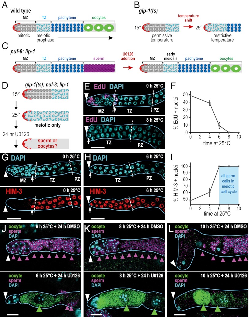
The mitosis–meiosis and sperm–oocyte cell fate decisions are separable. (A) Organization of WT C. elegans adult germ line. The stem cell niche (red) resides at the distal end; mitotic germ cells (gray) occupy the MZ; germ cells then enter the meiotic cell cycle and progress through early meiotic prophase (aqua-colored crescents) in the TZ and through pachytene (blue) in the pachytene zone (PZ). XX adult hermaphrodites make only oocytes (green). (B) Distal end of glp-1(ts) adult gonad. Germ cells are in the mitotic cell cycle at permissive temperature (15 °C) but enter the meiotic cell cycle when shifted to restrictive temperature (25 °C). (C) XX puf-8;lip-1 mutants make only sperm (purple), but treatment with the MEK inhibitor U0126 induces functional oocytes. (D) Experimental design to test separation of mitosis–meiosis and sperm–oocyte decisions. glp-1(ts);puf-8;lip-1 triple mutants are shifted to restrictive temperature (25 °C) to drive all germ cells into meiotic prophase (aqua-colored crescents); then U0126 or a DMSO solvent control is added, and germ lines are scored 24 h later for sperm or oocytes. (E, G, H, and J–L) Extruded gonads outlined in light blue; white arrowhead marks distal end; white dashed lines mark zone boundaries; small white arrow marks most distal meiotic prophase crescent. (Scale bars, 10 μm.) (E) At 15 °C (Upper), EdU labeling reveals S-phase nuclei in MZ, and DAPI shows meiotic prophase crescents, beginning at row 10. After 8 h at 25 °C, EdU incorporation ceases and crescent-shaped nuclei extend to distal end (Lower). (F) EdU incorporation as a function of time at 25 °C. Few S-phase cells remain at 6 h, and none remain at 8 h. Plot shows means ± SEM. (G) At 15 °C, the MZ lacks HIM-3 and crescents begin at row 10. (H) At 6 h after the shift to 25 °C, the MZ has been replaced by HIM-3+ meiotic cells that form crescents. (I) Percentage of HIM-3+ cells present in the distal germ line (i.e., MZ plus distal TZ) as a function of time shifted to 25 °C; light blue marks when all germ cells have entered the meiotic cell cycle. Points show means ± SEM. (J–L) Gamete sex can be reprogrammed from sperm to oocyte in meiotic germ cells. Sperm were visualized with sperm-specific marker SP56 (purple arrowheads); oocytes were seen with oocyte-specific marker OMA-2 (green arrowheads). % oo+, percentage of oocyte-positive animals. (J) Animals shifted to 25 °C for 6 h (U0126, 42% oocyte-positive animals, n = 12; DMSO, 0% oocyte-positive animals, n = 17); (K) animals shifted to 25 °C for 8 h (U0126, 52% oocyte-positive animals, n = 27 germ lines; DMSO, 0% oocyte-positive animals, n = 20); and (L) animals shifted to 25 °C for 10 h (U0126, 44% oocyte-positive animals, n = 32; DMSO, 4% oocyte-positive animals, n = 27 germ lines).

