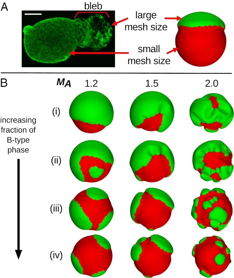
Fig. 1.
Relating mesh size in (A) experimental and simulation structures along with (B) simulation results for different parameters. (A) Nucleus from a breast cancer cell with lamin A stained, showing a larger mesh size in the bleb compared with the rest of the nucleus. (Scale bar: 5 μm.) The A-type component in the simulations (green) is analogously assigned to have a larger mesh size than the B-type component (red). (B) Low-energy configurations for lamin meshwork systems with phase fractions of the red B-type component of (i) f = 0.2, (ii) f = 0.4, (iii) f = 0.6, and (iv) f = 0.8. From left to right, the columns show systems with mesh size area scaling factors of MA = 1.2, 1.5, and 2.0, respectively, corresponding to the A-type component preferring a mesh size that is 20%, 50%, or 100% larger than the initial sphere, respectively. For clarity, dual meshes are shown here rather than vertices.

