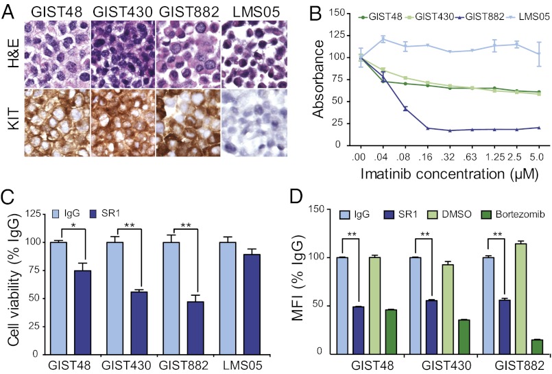
Fig. 1.
SR1 treatment slows in vitro GIST cell growth and reduces cell-surface KIT expression. KIT protein expression was analyzed by IHC on paraffin-embedded pellets of GIST48, GIST430, GIST882, and LMS05 cell lines (A). Cell viability assays were carried out to ascertain sensitivity to imatinib treatment for 72 h with GIST and LMS cell lines (B). Viable cell number, as measured by WST-1 absorbance, after 9 d in the presence of 10 μg/mL IgG control or SR1 was evaluated in GIST and LMS cells (C). Cell-surface KIT expression was evaluated in GIST cells by flow cytometry after 12 h of incubation with 10 μg/mL IgG, 10 μg/mL SR1, DMSO (1:1,000), or 100 nM bortezomib (D). All experiments were performed in triplicate. *P < 0.05 and **P < 0.01, as calculated by Student t test.

