Fig. 2.
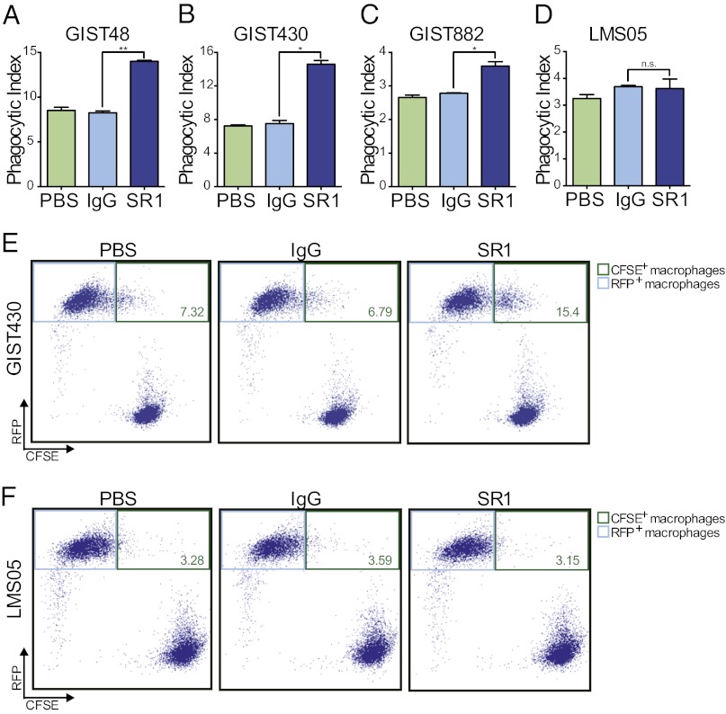
SR1 treatment enables GIST cell phagocytosis by macrophages. Macrophage phagocytosis of GIST and LMS cells was evaluated in the presence of PBS, 10 μg/mL IgG, or 10 μg/mL SR1 by flow cytometry. Macrophages that had successfully phagocytosed tumor cells were defined as RFP- and CFSE-double–positive cells (phagocytic index). SR1 treatment led to a significant increase in macrophage phagocytosis of GIST48 (A), GIST430 (B), and GIST882 (C) cells compared with IgG and PBS controls. SR1 treatment had no effect on macrophage phagocytosis of LMS05 cells (D). Representative dot plots are shown for GIST430 (E) and LMS05 (F). Upper right gates (green outline) were drawn to identify double-positive populations. The percentages of double-positive cells are labeled in green. All experiments were performed in triplicate. *P < 0.05, **P < 0.01, and n.s.P > 0.05, as calculated by Student t test.

