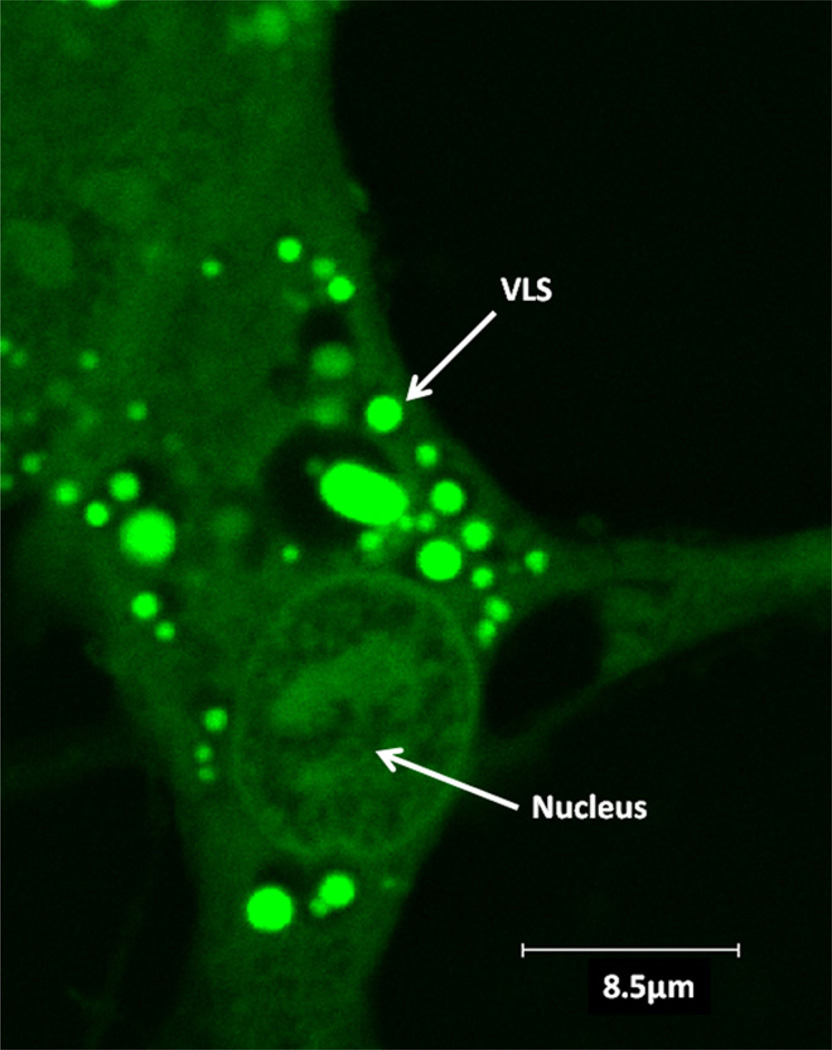
Fig. 1.
Periodontal fibroblasts treated with EMD–FITC and viewed by confocal laser scanning microscopy. A typical image of confluent HPDL fibroblasts incubated in culture for 17 h with 0.5 mg/ml EMD–FITC and viewed in monolayer by confocal laser scanning microscopy. Multiple, strongly fluorescent vesicle like structures (VLSs) were observed within the cytoplasm of the cells. Some vesicles exhibited a dark non florescent area surrounding a fluorescent central region.

