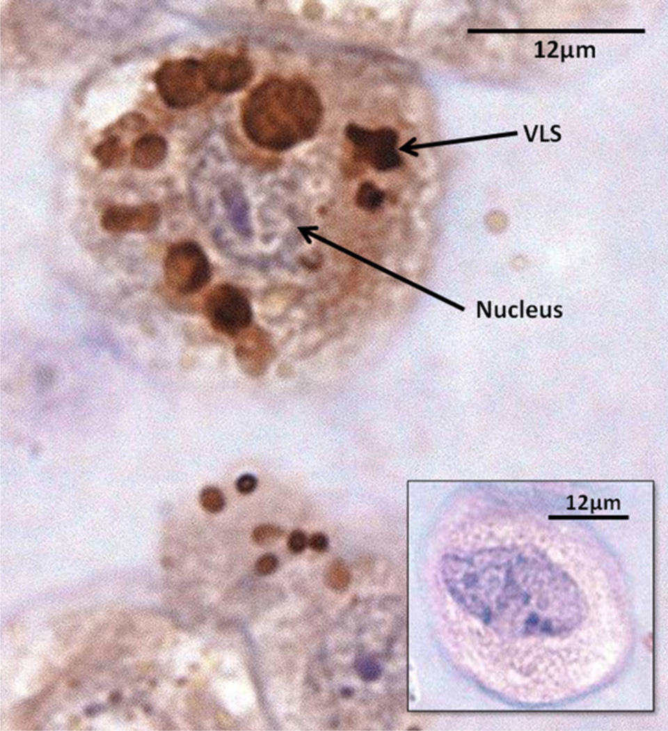
Fig. 2.
Paraffin sections of EMD–FITC treated HPDL fibroblasts probed with anti-20 kDa-amelogenin antibodies. Cells were counterstained with haematoxylin and eosin. Multiple, strongly cross-reactive VLSs were evident within the cytoplasm (arrowed). Inset shows negative control (no primary antibody). No significant cross-reactivity observed.

