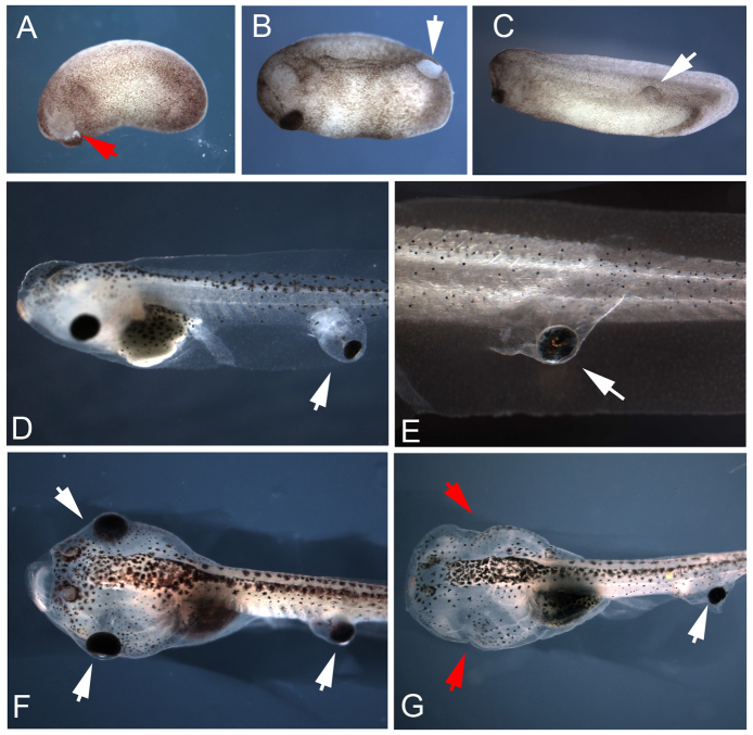
Fig. 1.
Eye primordia transplants produce ectopic eyes at the site of graft in Xenopus tadpoles. Donor tissue was excised from the eye field of stage 24 embryos (A, red arrow), grafted along the recipient's body, and healed within 30 min (B, white arrow). Ectopic eyes develop at the same rate as native eyes, and wounds heal completely within 24 h post-surgery (C). Eyes transplanted to caudal regions develop within pockets of tissue (D) or tightly along the trunk of the tail (E). Tadpoles receiving grafts could also have their native eyes removed through surgery at stage 46 (F, red arrows in G), leaving only ectopic visual structures.

