
Figure 2. MMP3 was overexpressed in BAVM.
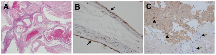
A, HE staining (magnification×40) verified a typical histological character of BAVM. B, the immunohistochemical staining showed that MMP3 was localized mainly in the endothelial cell layer of BAVMs (arrows, magnification×200). C, MMP3 was strongly immunostained in neurons (triangle) but not in vascular structures (arrows, magnification×400).