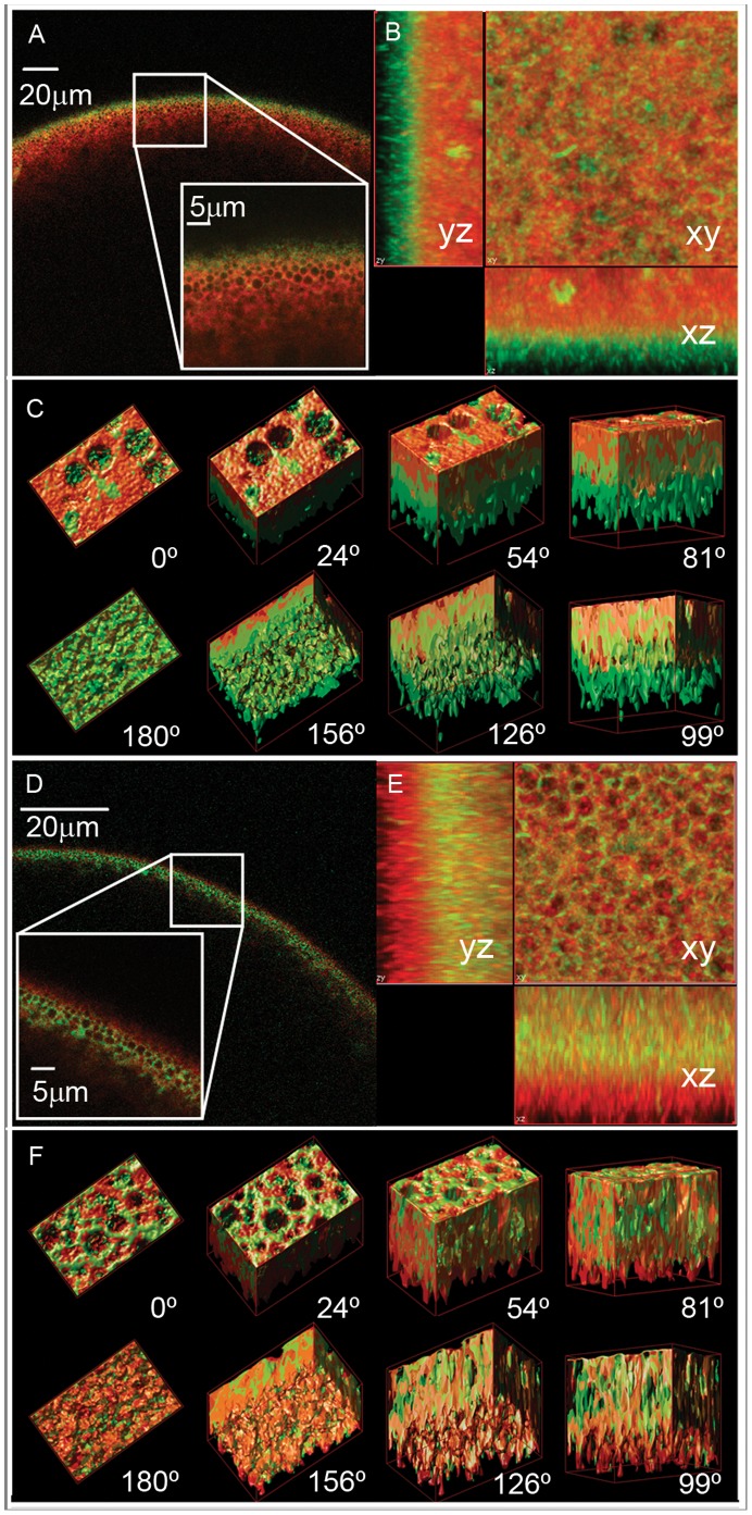
Figure 3. Localization of BvPIP1;1-ECFP when co-expressed with BvPIP2;2 or BvPIP2;1 in Xenopus laevis oocytes.
Radial (x–z) confocal images of X. laevis oocytes co-expressing BvPIP1;1-ECFP:BvPIP2;2 (green) (A) or co-expressing BvPIP1;1-ECFP:BvPIP2;1 (green) (D), both previously injected with TMR-Dextran (red). The oocyte surface is near the top of each image frame and the interior of the oocyte is in the bottom. Inside the image the enlargement of the indicated square section is shown. Stack of confocal (x–y) images were collected at various focal depths into the oocyte and then deconvolved and surface-render reconstructed with Huygens Professional Software. (B) Projections of the z-stack of images acquired with 100 nm step for oocytes co-expressing BvPIP1;1-ECFP:BvPIP2;2 (green) and (E) for oocytes co-expressing BvPIP1;1-ECFP:BvPIP2;1(green). (C) and (F) shows several views of the 3D reconstructed images for oocytes co-expressing BvPIP1;1-ECFP:BvPIP2;2 (green) and BvPIP1;1-ECFP:BvPIP2;1(green), respectively. The 0° view corresponds to the cortical granules level inside the oocyte and 180° to the plasma membrane plane (approximately 5 µm from the cortical granules level).

