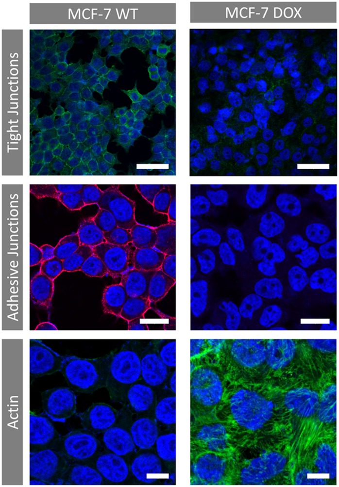
Figure 4. Immunocytochemical staining of cellular tight junctions (bar = 50 µm, green: occludin-Cy-2, blue: DAPI), adhesive junctions (bar = 20 µm Red: E-cadherin - Rhodamine, blue: DAPI) and actin (bar = 10 µm, green: Phalloidin-FITC, blue: DAPI) for MCF-7 WT and MCF-7 DOX cells.
Loss of tight junctions and adhesive junctions is observed upon gaining drug resistance. No E-cadherin was identified for MCF-7 DOX cells. Drug resistant cells displayed a highly dense fiber-like network of actin filaments and more cell-to-cell contact compared to MCF-7 WT cells.

