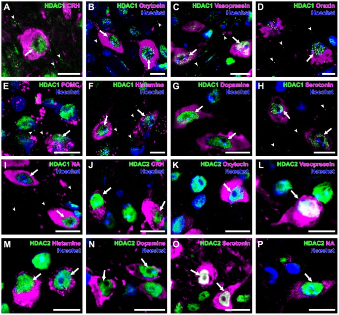
Figure 1. Expression of HDAC1 and -2 in monoaminergic and neuropeptidergic neurons.
The HDAC immunolocalizations (green) were visualized with a neuronal marker (magenta) and Hoechst stain (blue). A: HDAC1 was detected in the nucleus (arrow) of a CRH neuron, with punctate immunoreactivity (arrowheads) in the PVN. B: HDAC1 was detected in the nuclei (arrows) of oxytocin neurons, with punctate immunoreactivity (arrowheads) in the PVN. C: HDAC1 was detected in the nuclei (arrows) of vasopressin neurons, with punctate immunoreactivity (arrowheads) in the PVN. D: HDAC1 was detected in the nucleus (arrow) of orexin neuron, with punctate immunoreactivity (arrowheads) in the LHA. E: HDAC1 was detected in the nucleus (arrow) of POMC neurons, with punctate immunoreactivity (arrowheads) in the ARC. F: HDAC1 was detected in the nuclei (arrows) of histamine neurons, with punctate immunoreactivity (arrowheads) in the TMN. G: HDAC1 was detected in the nuclei (arrows) of dopamine neurons. H: HDAC1 was detected in the nuclei (arrows) of serotonin neurons, with punctate immunoreactivity (arrowheads) in the DR. I: HDAC1 was detected in the nuclei (arrows) of noradrenaline neurons, with punctate immunoreactivity (arrowheads) in the LC. J: HDAC2 was detected in the nuclei (arrows) of CRH neurons. K: HDAC2 was detected in the nucleus (arrow) of oxytocin neuron. L: HDAC2 was detected in the nucleus (arrow) of vasopressin neuron. M: HDAC2 was detected in the nuclei (arrows) of histamine neurons. N: HDAC2 was detected in the nuclei (arrows) of dopamine neurons. O: HDAC2 was localized in the nuclei (arrows) of serotonin neurons in the DR. P: HDAC2 was detected in the nucleus (arrow) of noradrenaline neuron. Scale bars indicate 10 µm.

