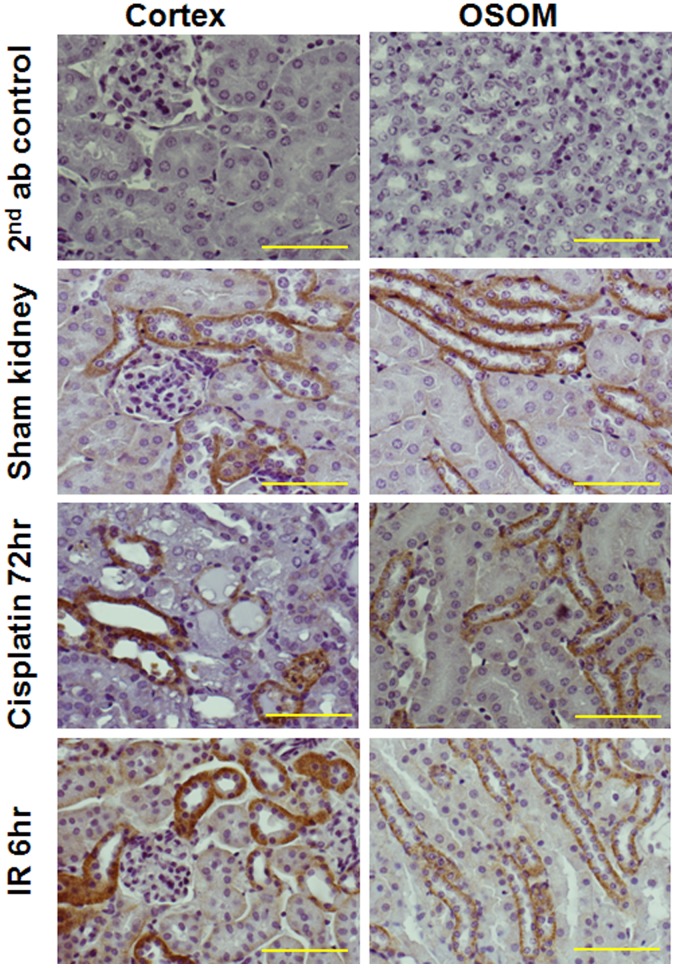
Figure 1. Immunohistochemical localization of semaphorin 3A in kidney after different treatments.
Immunohistochemical localization of semaphorin 3A was carried out as described in Materials and Methods. Immunostaining for semaphorin 3A is seen in thick ascending limb of the loop of Henle and distal tubular epithelial cells. Scale Bar: 100 µM.

