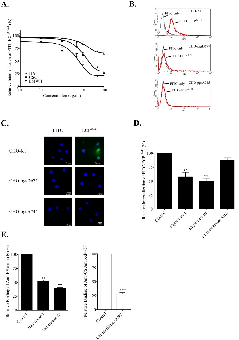
Figure 3. HS-dependent ECP32–-41 internalization.
(A) Beas-2B cells were treated with the indicated concentrations of LMWH, CS, or HA for 30 min prior to incubation with 5 µM FITC-ECP32–41 at 37°C for 1 h. The cells were washed twice with 500 µl PBS, trypsinized at 37°C for 15 min, suspended in 500 µl PBS, and subjected to flow cytometry. The result is expressed as the mean ± S.D., n = 3. (B) Samples of wild-type and mutant CHO cells were each incubated with 5 µM FITC-ECP32–41 at 37°C for 1 h and then subjected to flow cytometry. (C) Samples of wild-type and mutant CHO cells were incubated with 5 µM FITC-ECP32–41 at 37°C for 1 h., then washed twice with 1 ml PBS, and fixed for CLSM. Nuclei were stained with Hoechst 34850. Scale bar: 20 µm. (D) Beas-2B cells were treated with heparinase I, heparinase III, or chondroitinase ABC for 2 h prior to incubation with 5 µM FITC-ECP32–41 at 37°C for 1 h. The cells were washed twice with 500 µl PBS, trypsinized at 37°C for 15 min, suspended in 500 µl PBS, and subjected to flow cytometry. Untreated cells served as the controls. The fluorescence of cells treated with FITC-ECP32–41 was set to 100%. The result is expressed as the mean ± S.D., n = 3. **, P<0.01. (E) Beas-2B cells were treated with heparinase I, heparinase III, or chondroitinase ABC for 2 h. After stained with anti-HS or anti-CS monoclonal antibodies, washed twice with 500 µl PBS, and hybridized with FITC-conjugated anti-mouse secondary antibody, cells were suspended in 500 µl PBS and subjected to flow cytometry. Untreated cells served as the control. The fluorescence of the untreated cells was set to 100%. The result is expressed as the mean ± S.D., n = 3. **, P<0.01 and ***, P<0.001.

