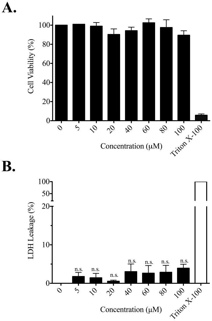
Figure 5. Cytotoxicity and membrane disruption by ECP32–41.
Beas-2B cells were grown in serum-free medium in the presence of ECP32–41 at indicated concentrations for 24 h. (A) The cytotoxic effect of ECP32–41 was measured by MTT assay. The cell viability of untreated cells was set to 100%. Cells treated with 0.1% Triton X-100 was used as a positive control. (B) The membrane disruption by ECP32–41 was measured by LDH assay. LDH released from cells lysed with 0.1% Triton X-100 in medium was defined as 100% leakage and LDH released from untreated cells was set as 0% leakage. The result is expressed as means ± S.D., n = 3. no significance (n.s.).

