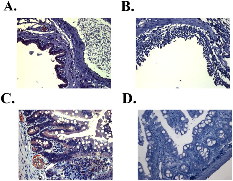
Figure 7. Tissue-specific localization of ECP32–41.
Immunohistochemical staining of ECP32–41 was detected by Supersensitive Non-Biotin HRP Detection System. Representative images of eGFP-ECP32–41 (red) in (A) broncho-epithelial and (C) and the epithelium of intestinal villi 1 h after intravenous injection of eGFP-ECP32–41 were shown. Signals of eGFP were not detected in (B) broncho-epithelial and (D) intestinal villi tissue sections 1 h after intravenous injection of eGFP. (Magnification in all panels, 200×).

