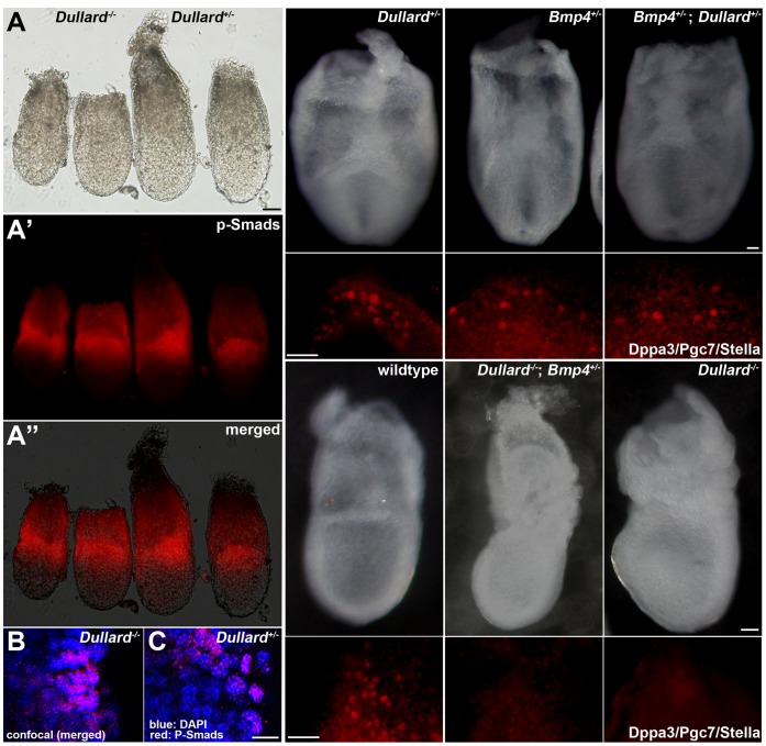
Figure 3. Dullard does not affect BMP4 signalling.
(A–A′′) The expression pattern of p-Smads in the proximal epiblast and adjacent extraembryonic ectoderm of the E6.75 Dullard −/− embryo was similar to that in Dullard +/− embryos (A, whole embryo bright-field view; A′, immunofluorescence image; A′′, merged image; left pair, Dullard −/− embryos; right pair, Dullard +/− embryos). (B, C) Confocal images of immunostaining of p-Smads in Dullard −/− and Dullard +/− embryos, respectively. (D–I′) Formation of Dpp3/Pgc7/Stella-positive PGCs was unaffected by the combined loss-of-function of Dullard and Bmp4. (D–F′) E7.75 compound heterozygous Dullard; Bmp4 embryos contained similar numbers of PGCs as those in Dullard+/− and Bmp4+/− embryos. (G–I′) Loss of Bmp4 activity in addition to complete loss of Dullard function had no effect on the PGC phenotype of E7.75 Dullard −/− embryos. (D–I) Whole embryo bright-field image; (D–G, posterior view; H, I, lateral views). (D′–I′) Dppa3/Pgc7/Stella immunostaining of the embryo in panels D–I. Scale bars = 100 µm (A–A′′), 20 µm (B, C) and 50 µm (D–I′).

