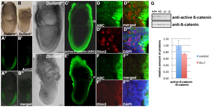
Figure 4. Down-regulation of WNT/β-catenin signalling activity in Dullard −/− embryos.
(A–B′′) Curtailed expression of Axin2 in the (B–B′′) E7.5 Dullard −/− embryo compared with that in the (A–A′′) Dullard +/− embryo. (A, B, bright-field images; A′, B′, Axin2 immunostaining; A′′, B′′, merged images). (C–F′′′) Expression of activated β-catenin (ABC) visualized by immunostaining and confocal microscopy of (C–D′′′) E7.5 no bud-stage Dullard +/− embryos and (E–F′′′) E7.5 Dullard −/− embryos. ABC expression in the posterior germ layer of the embryo, where Ifitm3-positive PGCs were localized, was weaker in the Dullard−/− embryo that also lacked Ifitm3-postive PGCs. (C, E) Bright-field image; (C′, E′) ABC immunostaining; (D–D′′′, F–F′′′) magnified views of the boxed areas in panels C′ and E′, respectively; (D′′, F′′) merged ABC and Iftm3 images; (D′′′, F′′′) merged images with DAPI nuclear staining; ABC immunostaining (green); Ifitm3 immunostaining (red), Scale bar = 100 µm (A–C′, E–E′) and 20 µm (D–D′′′, F–F′′′). (G) Western blot analysis of E7.75 wild-type (+/+) and Dullard +/− (+/−) embryos showing reduced amounts of the active form of β-catenin in Dullard−/− (−/−) embryos, but total β-catenin content was unchanged.

