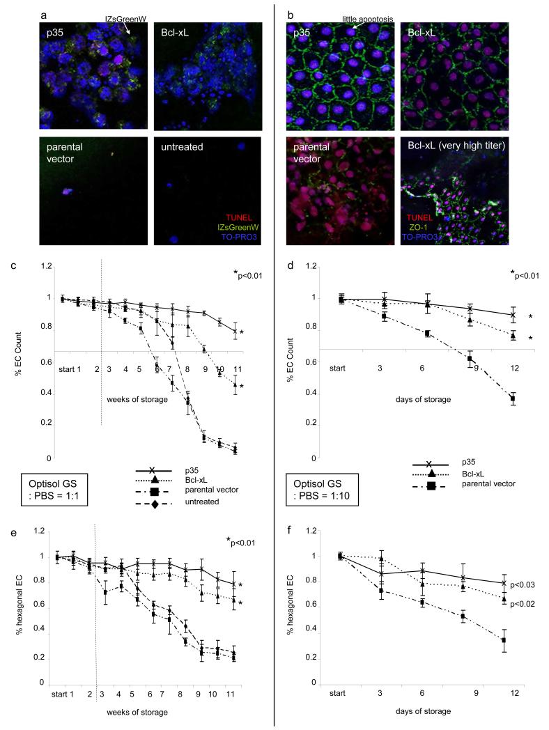
Figure 3. Assessment of corneal endothelial cell density and morphology during hypothermic storage at 4°C.
Untreated EC were compared to those expressing an parental vector (3 × 10^5 IU/ml), Bcl-xL (3 × 10^5 IU/ml) or p35 (3 × 10^5 IU/ml). EC densities and morphology of untreated endothelial cells and those expressing p35, Bcl-xL or an parental vector after storage for 11 weeks (a: Optisol GS:PBS=1:1) or for 12 days (b: Optisol GS:PBS=1:10) are demonstrated. Immunocytochemistry imaging in (a) and (b): TO-PRO3 (blue, nuclei), TUNEL (red, DNA fragmentation), IZsGreenW (a, green, co-expressed with the gene of interest) or ZO-1 (b, green, zonula occludens antibody). Endothelial cell count was enumerated in EC stored in diluted Optisol GS as indicated above. The point of interception of x- and y-axis in (c) and (d) corresponds to 2000 EC/mm2, the minimum EC count used by eye banks to release donor corneas for transplantation. Corneal endothelial cell morphology was evaluated by enumeration of hexagonal EC (e. Optisol GS:PBS=1:1, f. Optisol GS:PBS=1:10; the higher this ratio the faster the decrease of physiological hexagonality and EC numbers; six analyzed visual fields at each time point). The dotted black line in c and e indicates a period of 14 days during which the use of Optisol GS, the most widespread culture medium for hypothermic corneal storage, is currently accepted. P-values are relative to untreated controls only at the final timepoints (* = p<0.01).

