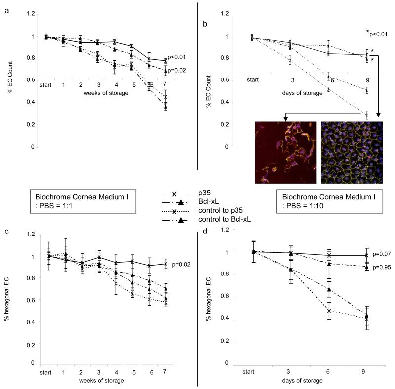
Figure 5. Expression of anti-apoptotic proteins leads to extended EC survival comparing paired corneas from the same donors cultured under conditions alike in an eye bank.
Organ storage in Biochrome Culture Medium I (1:1 or 1:10 dilution with PBS) was performed in donor pairs. One cornea of a pair was transduced with lenti-IZsGreenW, the other cornea with the lenti-IZsGreenW-Bcl-xL or lenti-IZsGreenW-p35. Enumeration of corneal endothelial cells is demonstrated in (a) and (b). The point of interception of x- and y-axis in (a) and (b) corresponds to 2000 EC/mm2, the minimum EC count used by eye banks to approve donor corneas for transplantation. Representative images of EC layers expressing IZsGreenW or p35 are shown in the respective inserts (TUNEL, TO-PRO3, ZO-1 (green) staining). Corneal endothelial cell morphology was evaluated by enumeration of hexagonal EC (c. Biochrome Culture Medium I:PBS=1:1, d. Biochrome Culture Medium I:PBS=1:10; eight analyzed visual fields at each time point). P-values are relative to untreated controls only at the final timepoints (* = p<0.01).

