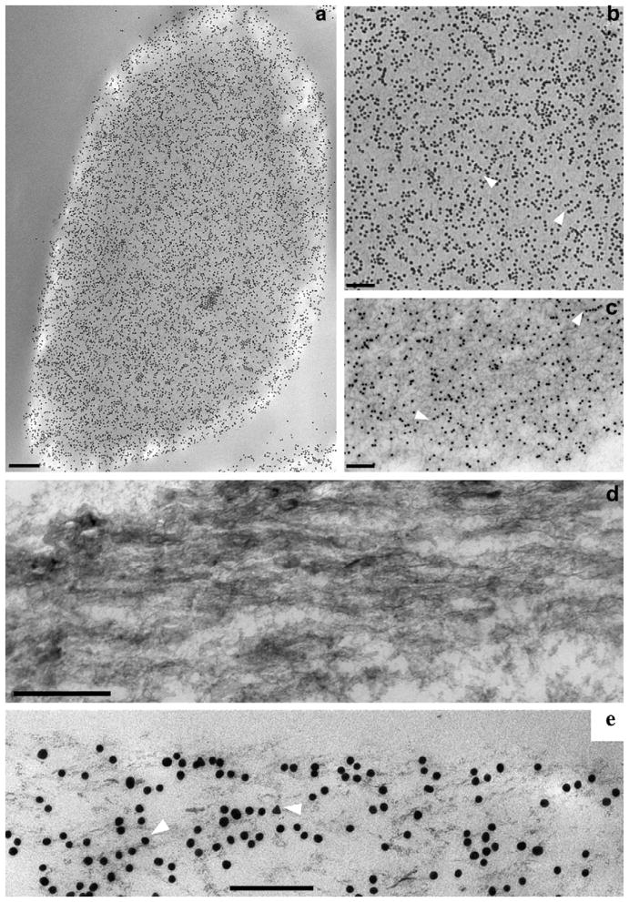
Fig 3.
Immunogold TEM of OC90 in outer cortex. (a) Low magnification view of longitudinal/tangential section of outer cortex, displaying high density of gold particles; (b) high magnification of Fig. 3a, demonstrating gold particles aligned in rows in straight or slightly curved patterns (arrowheads); (c) EDTA-treated cortical region of an otoconium, distinctly showing dense unstained fibrillar matrix as well as immunogold particles. In most regions gold particles are aligned in same direction as fibrils (arrowheads); (d) ultra-thin sagittal section of outer cortex of mouse otoconium; (e) corresponding section, immunogold labeled for OC90, indicating that the majority of the particles are aligned in the same direction of mineralized fibrils shown in (d) (white arrowheads). Bars = 0.15 μm (a); 0.2 μm (b); 0.2 μm (c); 0.1 μm (d); 0.1 μm (e).

