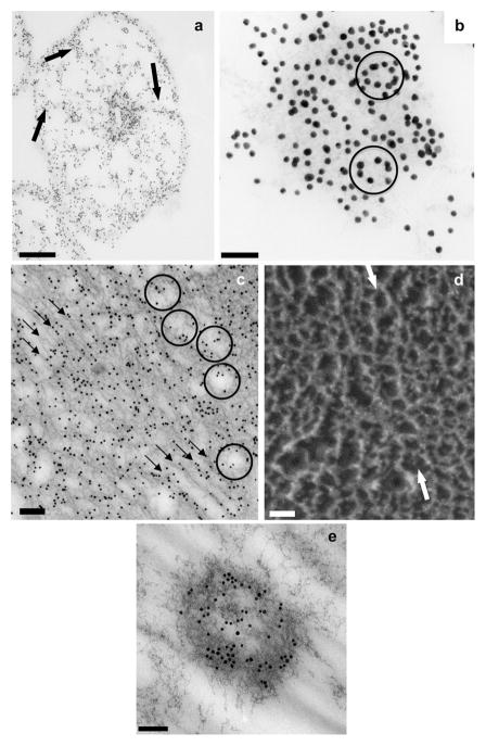
Fig 4.
Immunogold TEM of OC90 and deep-etch images of otoconia. (a) Cross-section through the equatorial plane of mouse otoconium showing strongly labeled inner core. Thin spoke-like projections radiate to periphery, merging with densely labeled subsurface layer (arrows); (b) high magnification image of central core showing several oval gold particle alignments (arrows) in addition to straight or slightly curved patterns; (c) longitudinal section of an EDTA-treated gold labeled inner core. Because of EDTA treatment, the unstained matrix is sharply contoured. Three rows of gold particles in the center of image delineate sets of diagonally oriented tubular formations corresponding to cavities in the fibrillar scaffold. Several diagonally oriented contiguous, co-aligned rows of oval/polygonal fibrillar formations are seen in periphery of image (arrow); (d) deep-etch image of freeze-fractured inner core, showing several rows of oval, co-aligned fibrillar formations (arrows) analogous to those seen in the thin sectioning TEM of Fig. 4c. [Note that two totally different techniques (thin sectioning TEM and freeze fracturing) show identical patterns.] (e) ectopic immunogold labeled structure resembling inner core on an otoconium, located in gelatinous membrane. Bars = 1 μm (a); 0.05 μm (b); 0.125 μm (c); 0.05 μm (d); 0.1 μm (e).

