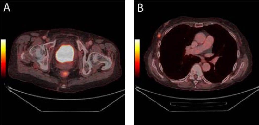
Figure 1.
Muscle invasive bladder cancer in a 78-year-old patient. The bladder tumour is not visible on axial 18F-FDG PET/CT images due to urinary excretion of 18F-FDG which mask the tumour (1A). No metastases from the bladder tumour were demonstrated. However, a mass suspicious for malignancy was demonstrated in the right breast as can be seen on axial fused PET/CT images (1B). Histology confirmed a primary breast cancer.

