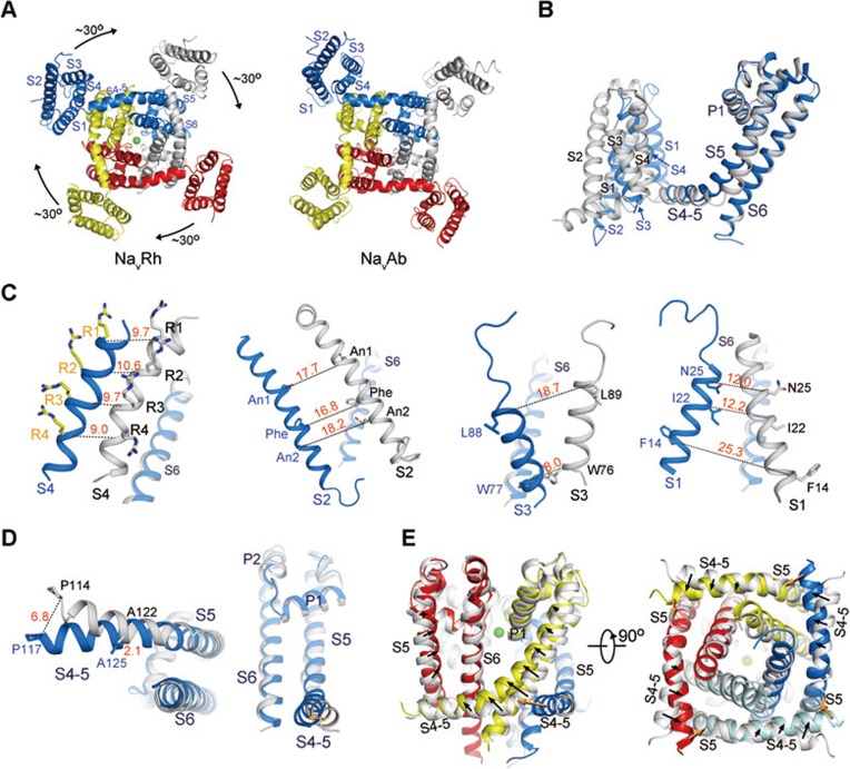
Figure 1.
The conformational shifts between NavRh and NavAb. (A) Lateral rotation of the VSDs between NavRh and NavAb when the pore domains are superimposed. A cytoplasmic view is shown. Please refer to Supplementary information, Movie S1 for a morph of the overall conformational changes between NavRh and NavAb. (B) Structural changes between individual protomers when the structures of the tetrameric NavRh and NavAb are superimposed relative to the pore domains. NavRh and NavAb are colored blue and grey, respectively. (C) Dissection of the conformational shifts of the VSD segments relative to the pore domain. The S6 segments are shown in each panel as the reference to indicate the orientation of the structures. (D) The conformational shifts of the S4-5 helix as well as the S5 segment relative to the pore. A cytoplasmic view and a side view are shown. (E) The S4-5 helices and S5 segments are shifted (indicated by the black arrows) between NavRh and NavAb. The pore domain of NavRh is colored the same as in (C), and that of NavAb is colored silver. The orange arrows indicate the potential pushing force exerted from the adjacent protomer during the conformational changes of its VSD.

