Figure 1.
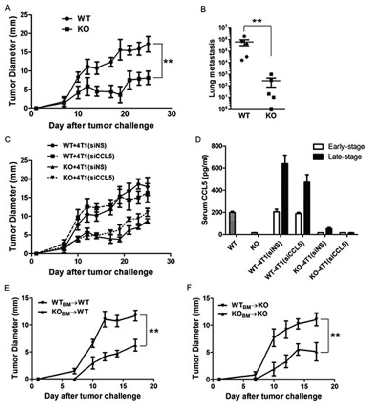
4T1 tumor growth and metastasis in CCL5 KO mice. (A) 4T1 tumor cells were injected into the mammary gland of female WT and KO mice. Tumor growth was monitored every 2-3 days. Data are represented as mean ± SE. Each group contained five mice. (B) Lung metastasis of 4T1 tumor cells was measured by the 6-thioguanine clonogenicity assay. When tumor diameter reached 20 mm, mice were sacrificed and the number of lung metastasis was quantified. (C) Tumor growth in WT and KO mice injected with control cell line 4T1(siNS) or 4T1(siCCL5). n = 5 mice per group. (D) 10 days (early stage) or 20 days (late stage) after tumor cell inoculation, mice were sacrificed and serum was collected for measurement of CCL5 protein by ELISA. n = 5 mice per group. (E, F) Lethally irradiated female WT and KO mice were transplanted with male WT (E) and KO (F) bone marrows. Thirty days after bone marrow reconstitution, the recipients were inoculated with 4T1 cells and tumor growth was monitored every 2-3 days. Data are represented as mean ± SE. Each group contained three mice. **P < 0.01.

