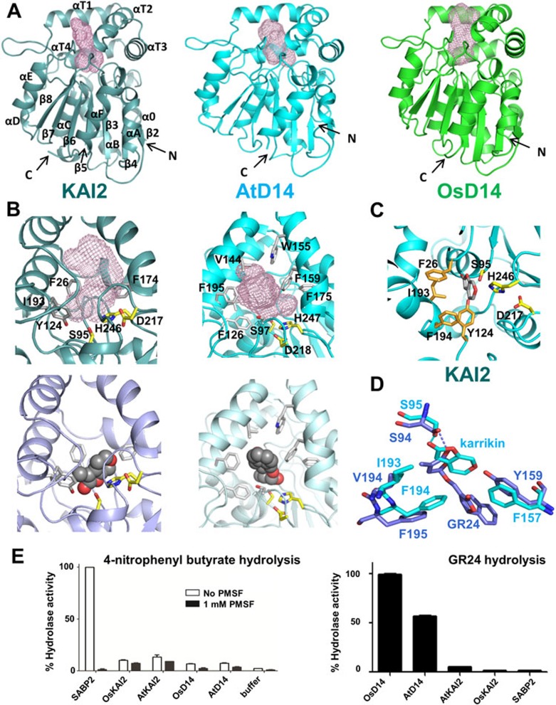
Figure 1.
Structures and activities of KAI2 and D14 proteins. (A) Structure overview of apo KAI2, AtD14, and OsD14. Ligand-binding pockets are indicated as mesh. (B) KAI2 and D14 ligand-binding pockets (top) and docked ligands (bottom). (C) Close-up view of the KAI2 catalytic triad with docked ligand. (D) Structural alignment of ligands and key ligand specificity-conferring residues. (E) Hydrolase activity of KAI2, D14, and SABP2 toward a generic small substrate (left) and GR24 (right).

