Figure 4.
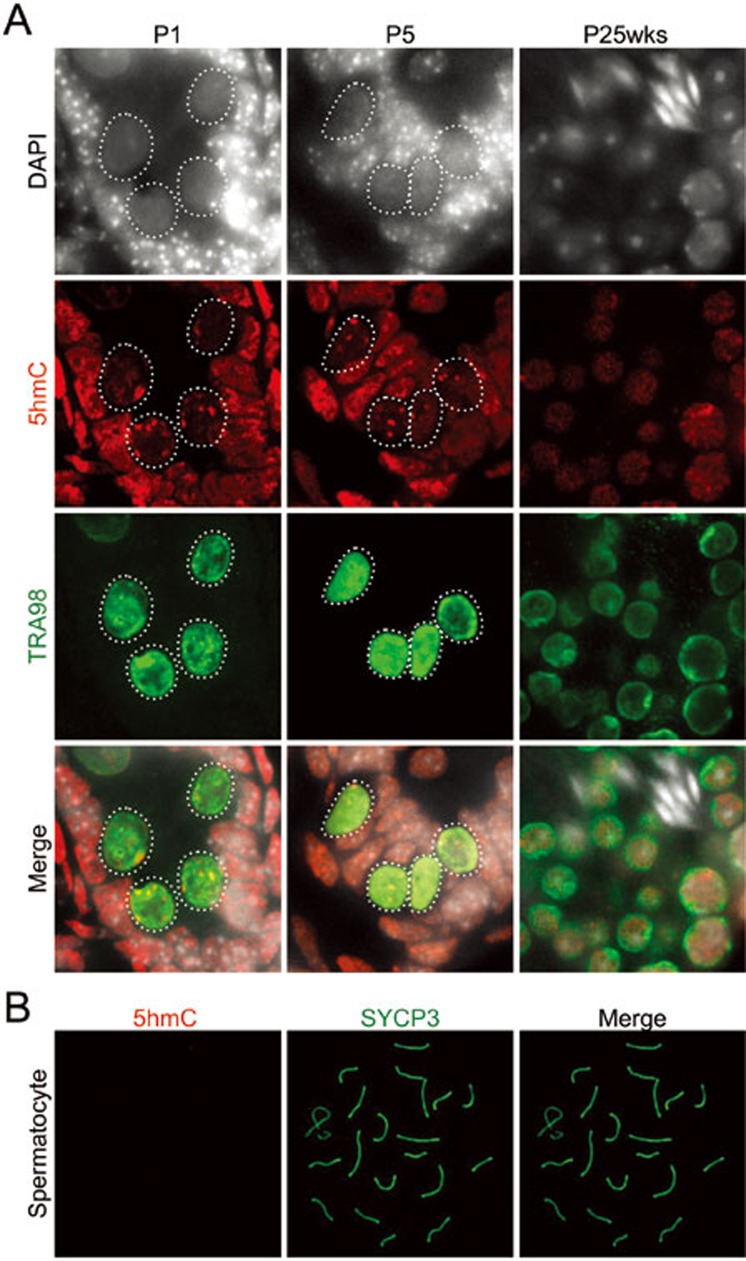
Loss of pericentric 5hmC during postnatal male germ cell development. (A) Representative images of cryosections of P1, P5, and 25-week-old testis co-stained with 5hmC (red) and germ cell marker TRA98 (green) antibodies. Dashed circles indicate germ cells. (B) Representative images of pachytene stage spermatocyte co-stained with 5hmC (red) and a synaptonemal complex marker SYCP3 (green). No 5hmC signal was detected at this stage.

