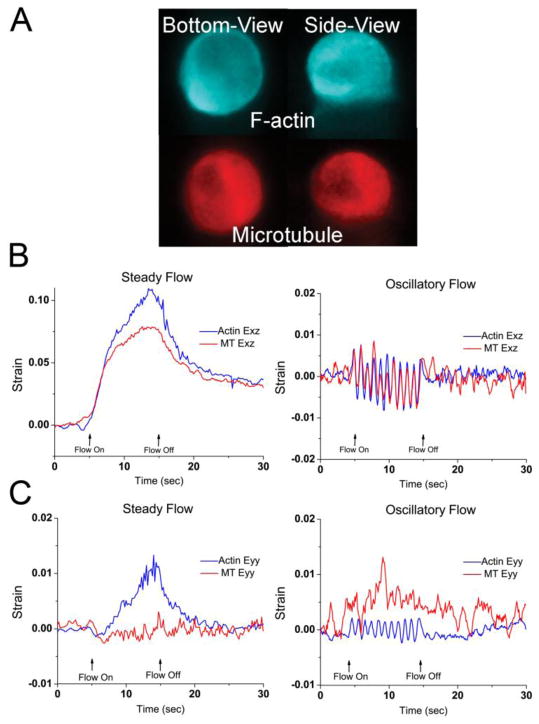
Figure 2.
A) Sample image of the F-actin and microtubule networks of an MLO-Y4 osteocyte imaged simultaneously in two orthogonal planes (bottom and side views). B) Sample side-view whole-cell shear Exz strain trace for both cytoskeletons under steady flow or oscillatory flow. Both networks display creep and creep-recovery behaviors in steady flow. Both networks display sinusoidal shear strains that follow the oscillatory flow pattern. C) Sample side-view whole-cell shear Eyy strain trace for both cytoskeletons of a single osteocyte under steady or oscillatory flow that show different mechanical responses within the same cells between the two cytoskeletons. In these examples, only the actin network displays characteristic creep or oscillation.

