Fig. 2.

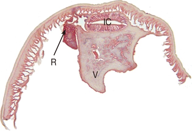
Transverse section of the P. decipiens larva. Note that the intestinal cecum (IC), ventriculus (V), and renette cell (R) are seen.

Transverse section of the P. decipiens larva. Note that the intestinal cecum (IC), ventriculus (V), and renette cell (R) are seen.