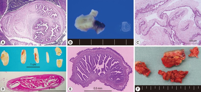
Fig. 2.
Histopathologic findings of cystic lesions from cysticercosis patients. (A) A cellulose-type cysticercus characterized by rabyrinth-like structure (case 40 [22], courtesy of Dr. S. Matsunaga). (B) and (E) A resected lesion and a cellulose-type cysticercus (case 48 [32], courtesy of Prof. H. Matsuoka). (C) Racemose-type cysticercus characterized by complicated cystic walls (case 62 [40], courtesy of Dr. S. Shiiki). (D) SCC showing typical rice grain calcifications in the muscles of buttocks and lower extremity and the section of the calcified lesion (case 44 [27, 28], courtesy of Dr. T. Nagase). (F) Surgically removed calcified lesions (case 52 [36], courtesy of Dr. Tsuda). Sections (A, C , D, and E) were stained with hematoxylin and eosin.

