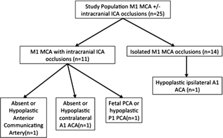
Figure 4.
Distribution of incomplete/hypoplastic circle of Willis in the study population stratified by site of occlusion. In 11 patients with intracranial internal carotid artery (ICA) occlusions, only 2 patients had an incomplete anterior circle of Willis as a potential explanation for dominant posterior cerebral artery (PCA)–middle cerebral artery (MCA) leptomeningeal collateral pattern. In 14 patients with isolated M1 MCA occlusions, only 1 patient had an hypoplastic ipsilateral A1 anterior cerebral artery (ACA) as a potential explanation for dominant posterior PCA–MCA leptomeningeal collateral pattern.

