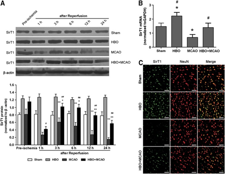
Figure 1.
Hyperbaric oxygen (HBO) preconditioning upregulates SirT1 protein and mRNA in ischemic penumbra of the brain. (A) Representative western blot bands and quantitative analysis of the time course of SirT1 protein expressions (n=5/time point/group). *P<0.05, **P<0.01 versus preischemia; #P<0.05, ##P<0.01 versus middle cerebral artery occlusion (MCAO). The expression of SirT1 was normalized to the expression of β-actin. (B) The expression of SirT1 mRNA was analyzed using quantitative real-time PCR 3 hours after reperfusion (n=5/group). *P<0.01 versus Sham; #P<0.01 versus MCAO. The expression of SirT1 was normalized to the expression of glyceraldehyde 3-phosphate dehydrogenase (GAPDH). Data are presented as mean±s.d., one-way analysis of variance (ANOVA) followed by the least significant difference (LSD) test. (C) Representative microphotographs showing the double immunofluorescence staining for SirT1 (green) and neuronal nuclei (NeuN; neuronal marker, red) to determine the cell identity for SirT1 expression in ischemic penumbra of brain at 3 hours after reperfusion (n=3/group). Scale bars=50 μm. MCAO: rats subjected to occlusion of middle cerebral artery for 120 minutes; HBO: rats subjected to HBO preconditioning (2.5 atmospheres absolute, 100% O2, 1 h/day, 5 days); HBO+MCAO: rats subjected to MCAO 24 hours after the end of HBO preconditioning. The color reproduction of this figure is available at the Journal of Cerebral Blood Flow and Metabolism journal online.

