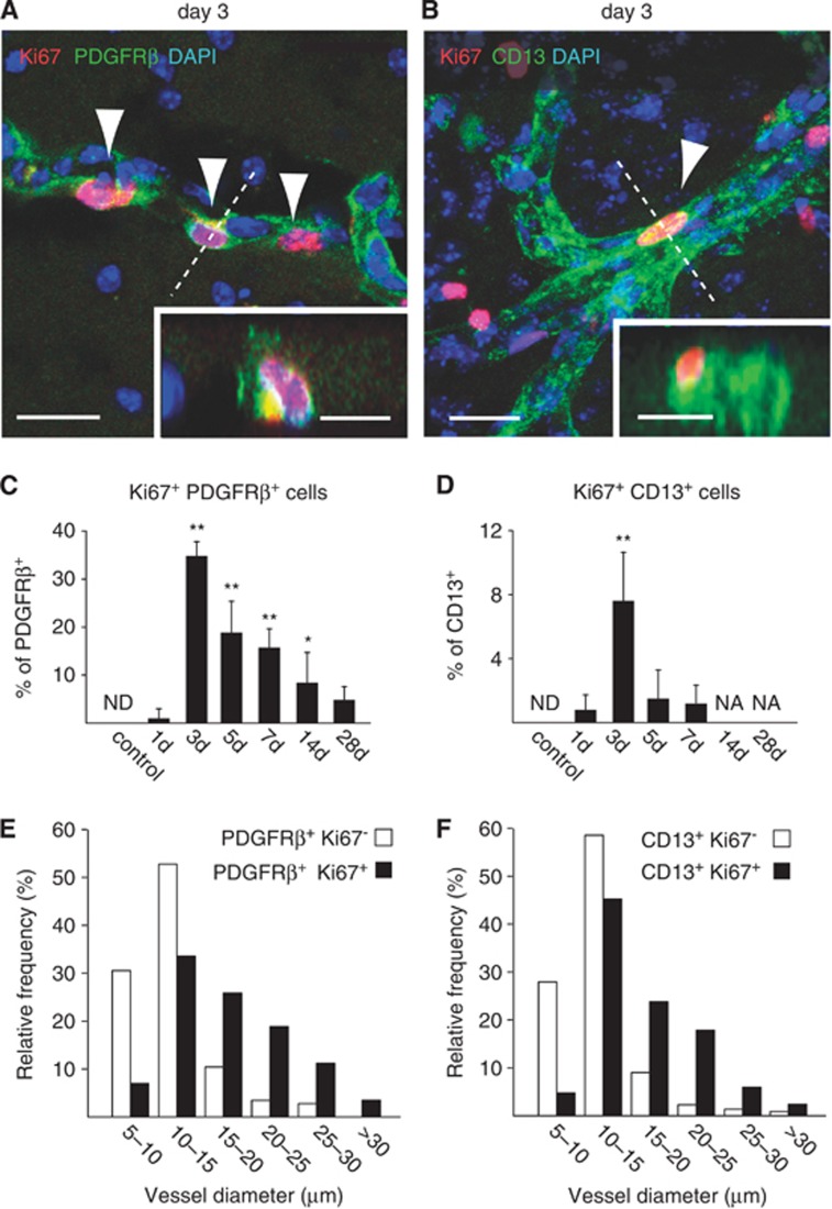
Figure 4.
Proliferation of platelet-derived growth factor receptor beta (PDGFRβ+) and CD13+ cells after middle cerebral artery occlusion (MCAO). (A, B) At day 3 after MCAO, Ki67 staining reveals proliferation of PDGFRβ+ cells (A) and CD13+ cells (B) at vessel walls (insets, orthogonal reconstructions from the planes indicated by dashed lines). (C, D) Quantification of Ki67-immunoreactive PDGFRβ+ cells (C) and Ki67-immunoreactive CD13+ cells (D) reveals significant bursts of proliferation at day 3 after MCAO. Whereas PDGFRβ+ cells still maintain robust proliferation at days 5 to 7, the proliferation of CD13+ cells subsides after day 3. (E, F) At day 3 after MCAO, frequency histograms of vessel size show that vessel-associated Ki67-immunoreactive PDGFRβ+ and CD13+ cells are predominantly found at microvessels with large diameters (>10 μm), rather than capillaries. Bars represent means±s.d. *P<0.05, **P<0.0005 versus control animals. n=5, 4, 4, 5, 3, 5, and 5 for control, 1, 3, 5, 7, 14, and 28 days, respectively. NA, not available; ND, not detected. Scale bars: 20 μm; 10 μm (insets).

