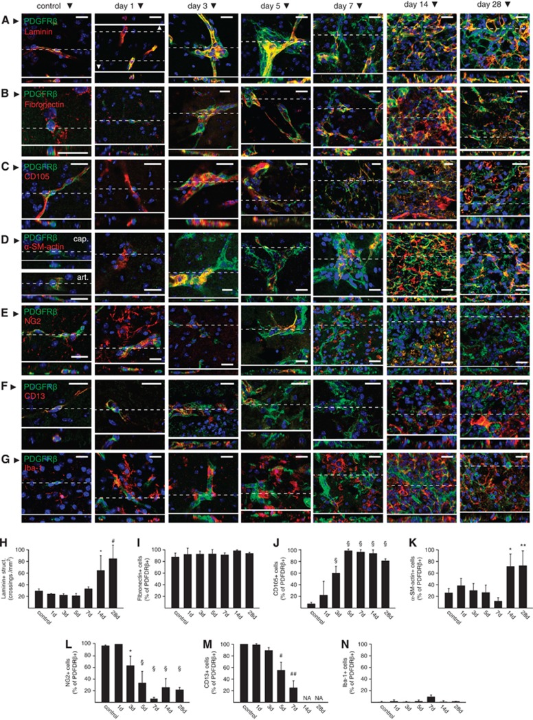
Figure 5.
Characterization of platelet-derived growth factor receptor beta (PDGFRβ+) cells in the ischemic brain tissue. (A–G) Representative examples showing single confocal planes of immunofluorescence stains for PDGFRβ and laminin (A), fibronectin (B), CD105 (C), α-smooth muscle (SM)-actin (D), NG2 (E), CD13 (F), and Iba-1 (G) in control tissue or at the core of ischemia at different time points after middle cerebral artery occlusion (MCAO). Orthogonal reconstructions of the confocal stacks (insets) obtained from the planes marked by dashed lines are shown for each figure. (H–N) Quantification of laminin immunoreactive extracellular matrix (ECM) structures (H) and the fraction of PDGFRβ+ cells immunoreactive for fibronectin (I), CD105 (J), α-SM-actin (K), NG2 (L), CD13 (M), and Iba-1 (N) in control tissue or in the core of ischemia at different time points after MCAO. Collectively, the data show the proliferation of laminin+ ECM in the core of ischemic tissue, gain of a stromal or myofibroblast phenotype by PDGFRβ+ cells (colocalization with fibronectin, CD105, or α-SM-actin), and loss of pericyte (PC)/mural cell markers (loss CD13 or NG2 immunoreactivity). Bars represent means±s.d. *P<0.005, **P<0.001, #P<0.0001, ##P<0.00001, §P<0.000001 versus control animals. n=5, 4, 4, 5, 3, 5, and 5 for control, 1, 3, 5, 7, 14, and 28 days, respectively. NA, not available. Scale bars: 20 μm.

