Figure 1.
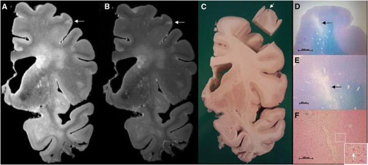
Sampling procedure: correlation of ex vivo magnetic resonance imaging (MRI) and histology. The sampling procedure was developed in three brain slices from anonymous donors. In one of these brain slices a cortical lesion of interest, presumed to be ischemic, was identified on ex vivo MRI. This lesion appeared as a hyperintense cortical lesion with a hypointense center on a fluid attenuated inversion recovery (FLAIR) (A; 400 × 400 × 400 μm3), and as a hyperintense cortical lesion on a T2 weighted (B; 400 × 400 × 400 μm3) magnetic resonance (MR) image. After dissection of the tissue for sampling a cavity in the cortex was visible (C; tissue block is turned over). On histopathologic examination, this cortical lesion was identified as a cerebral microinfarct (CMI) with cavitation and gliosis in the surrounding tissue (D, and in greater detail E) (Luxol fast blue & Periodic acid-Schiff (L&P) stain). On higher magnification, gliosis is visible (F; arrow indicates a reactive astrocyte) (hematoxylin/eosin (HE) stain). Tissue was submerged in Fomblin.

