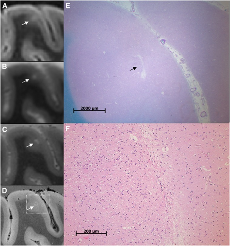
Figure 4.
A cerebral microinfarct (CMI) identified with ex vivo magnetic resonance imaging (MRI). A CMI in a formalin-fixed coronal brain slice of a 64-year-old patient with a neuropathologic diagnosis of Alzheimer's disease (AD). The CMI is presented as a hyperintense cortical lesion on a clinical resolution fluid attenuated inversion recovery (FLAIR) (A; 0.8 × 0.8 × 0.8 mm3) and a clinical resolution T2 weighted image (B; 0.7 × 0.7 × 0.7 mm3), and as a hyperintense cortical lesion in greater detail on an ultra-high resolution T2 weighted (C; 400 × 400 × 400 μm3) magnetic resonance (MR) image. On an ultra-high resolution T2* weighted (D; 180 × 180 × 180 μm3) MR image, the CMI can be observed in even greater detail and in close resemblance to the microscopic images in (E) and (F). On histopathologic examination, this cortical lesion was established as a CMI with moderate gliosis in the surrounding tissue (E; Luxol fast blue & Periodic acid-Schiff (L&P) stain, and in greater detail F; hematoxylin/eosin (HE) stain).

