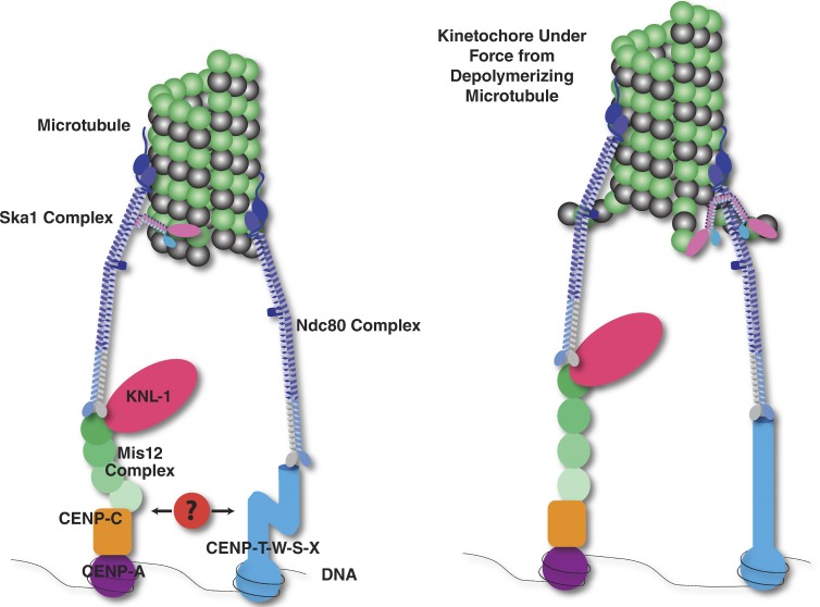
Figure 1.
Simplified diagram of the kinetochore showing the major proteins involved in the DNA–microtubule attachment. (Left) The Ndc80 complex (dark blue) binds to microtubules and forms two separate connections to kinetochores. First, the Ndc80 complex binds to the Mis12 complex (green) and KNL-1 (magenta). The Mis12 complex in turn binds to CENP-C (orange), which binds to nucleosomes containing the histone H3 variant CENP-A (purple). Second, the Ndc80 complex binds to CENP-T (light blue). CENP-T interacts with DNA as a part of a heterotetrameric nucleosome-like CENP-T–W–S–X complex. In humans, the Ndc80 complex attachment to microtubules is enhanced by an interaction with the Ska1 complex (pink and blue; Schmidt et al., 2012). Additional components may form interactions between the two connective pathways (red). (Right) Upon microtubule depolymerization, the flexible protein components of the kinetochore may rearrange. For example, recent evidence has suggested that the N and C termini of CENP-T separate under tension (Suzuki et al., 2011) and that the subunits of the Mis12 complex redistribute (Wan et al., 2009).

