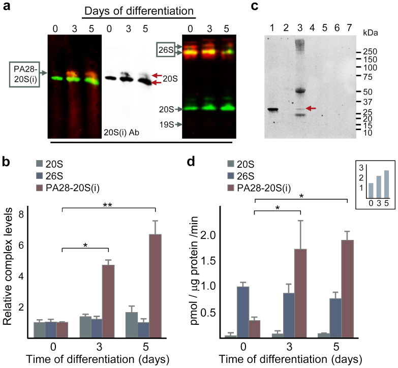
Figure 3. A functional proteasome activator PA28 is produced upon differentiation of ES cells.
(a) Levels of mature PA28-20S(i) (left; detected using antibodies against 20S α-subunits [green] and PA28α [red] on a blotted gel which had been run for 8.5 h; the middle blot displays the 20S channel alone) and 26S (right; detected using antibodies against the 19S subunit Rpn7 [red] and 20S α-subunits [green] on a blotted gel which had been run for 12 h) proteasomes. (b) Proteasome complexes PA28-20S(i) (red), 26S proteasome (blue), or 20S proteasome (grey) quantified from (a) and additional runs, error bars represent SD (n ≥ 2); p* = 0.013 and p** = 0.0040 (one-way ANOVA followed by Tukey's post hoc test, see Supplementary Fig. S8). (c) Immunochemical detection of PA28α before and after 20S immunoprecipitation (IP) of 3-day differentiated ES cells; lanes from the left: (1) untreated cell extract, (2) beads used to pre-clear extract, (3) 20S-IP beads (arrow indicates PA28α), followed by 4 lanes (4–7) of washes of the 20S-IP beads. (d) Proteasome activity measured under conditions optimizing for either PA28-20S(i) (red), 26S proteasome (blue), or 20S proteasome (grey) assembly and activity, error bars represent SD (n ≥ 2); p*day0-day3 = 0.037 and p*day0-day5 = 0.017 (one-way ANOVA followed by Tukey's post hoc test, see Supplementary Fig. S9). Inset shows the combined proteasome activity in pmol LLVY digested per μg protein and min, i.e. the sum of activity measurements favouring PA28-20S(i), 26S proteasome, and 20S proteasome after 0, 3, and 5 days of differentiation.

