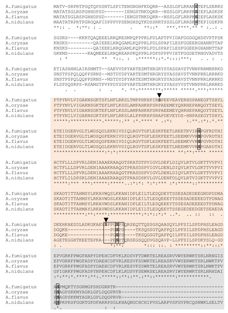
Fig. 3.
Clustal alignment of AspE homologs from other Aspergillus species showing conservation of phosphorylated residues. AspE homologs from other Aspergillus species were aligned using the ClustalW program. The sites of conserved phosphorylated residues are boxed and indicated in red color. The non-conserved phosphorylated residues are indicated by arrowheads. The short serine-proline rich region is boxed in red. The N terminal (in white) and C terminal domains (in grey) and the G-domain (in orange) are shown.

