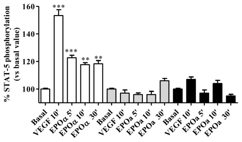
Figure 1.
Vascular endothelial growth factor (VEGF)- and epoetin alpha (EPOα)-mediated STAT-5 phosphorylation. Human umbilical vein endothelial cells (HUVECs) were treated with 25 ng/mL VEGF (10 min) or 1 IU/mL EPOα (5–10 and 30 min) under three different cell culture conditions. White bar: replacement of cell culture medium for 24 h with M199 without fetal bovine serum (FBS) and growth factors supplemented with 0.5% bovine serum albumin (BSA) and 0.2 mM orthovanadate. Gray bars: replacement of cell culture medium for 24 h with M199 without FBS and growth factors. Black bars: replacement of cell culture medium for 12 h with M199 medium without FBS and growth factors supplemented with 0.2 mM orthovanadate 30 min before the experiments. In each experimental condition, cell responsiveness was evaluated by assessing whether VEGF or EPOα stimulated STAT-5 phosphorylation. The data are expressed as the percent STAT-5 phosphorylation compared to the untreated control cells (set to 100%) and represent the mean ± SEM of three different experiments. *** p < 0.001; ** p < 0.01 vs. basal value.

