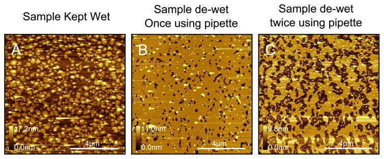
Figure 10.
Illustration of the dewetting test for DPPC. The sample bilayer in (A), which is very similar to the bilayer in Figure 9C, was dewetted instantaneously and immediately rehydrated (B); The dewetting step was repeated a second time (C). The holes in the bilayer become progressively larger after each successive wash. The test proves that the sample in (A) was a complete bilayer with partially fused lipid patches on top.

