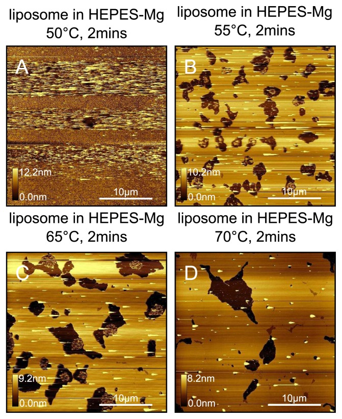
Figure 12.
DPPC bilayers prepared from a HEPES-NaCl-Mg liposome solution. Bilayers prepared at (A) 50 °C; (B) 55 °C; (C) 65 °C; (D) 70 °C. When the sample plate is maintained at 50 °C we only observe a vesicle layer, which can be partially fused by the tip as indicated by the horizontal streaks in A. For 55 °C, 65 °C and 70 °C deposition temperatures, we observe extended regions of fused bilayer. More domains are observed for the highest temperatures. Samples were prepared using liposomes in HEPES-Mg at exactly the same concentration (0.06mgml−1 ) and deposition time (2min) as for the HEPES-NaCl samples shown in Figure 11.

